Appointment Expectations
According to the American Medical Association, there are over 20,000 ophthalmologists in Salem and around the United States. An ophthalmologist is a physician that performs eye exams and surgeries. The difference between an ophthalmologist and an optometrist is that an ophthalmologist has additional years of study and experience treating the various conditions of the eye and can perform surgeries. Optometrists usually focus on routine eye health and basic vision needs, such as glasses or contact lenses.
When planning an appointment with your Salem ophthalmologist, it helps to know what to expect. There can be a lot going on and it can get confusing or overwhelming, so we’re going to walk you through everything you might encounter during your exam.
- Prep
- Staff
- Detection
- Machinery
- Testing
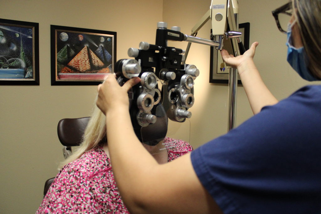
When the doctor enters the room, the scribe follows to document the doctor’s findings during the exam. The doctor begins the exam by making sure any questions the patient may have are addressed. The doctor will check each patient’s pupils and eye muscle movement for any abnormalities. Usually, the doctor will leave the room at this point to move on to his next patient. The scribe will stay behind and check the patient’s intraocular pressure and instill dilating drops. The dilating drops enlarge the pupils and allow the doctor to see to the back of the eyes. It takes about 20 minutes to get the full effect, and can last for a few hours. When the doctor returns he will preform a refraction, which new prescriptions for glasses are determined or serves as abase line vision for catalytic surgery.
The effects of dilation include trouble focusing up close and some light sensitivity. Once the pupils are fully dilated, the doctor uses the slit lamp microscope and different magnifying lenses to look at the eyes from front to back. During this examination is where the doctor would detect any conditions that may or may not need treatment. Some common ocular conditions are cataracts, diabetic retinopathy, macular degeneration, and glaucoma. Most of these diagnoses are initially made based on what the doctor sees during the exam. However, there are many tests we can perform to verify any findings that allow the doctor to further evaluate and manage each patient’s individual needs.
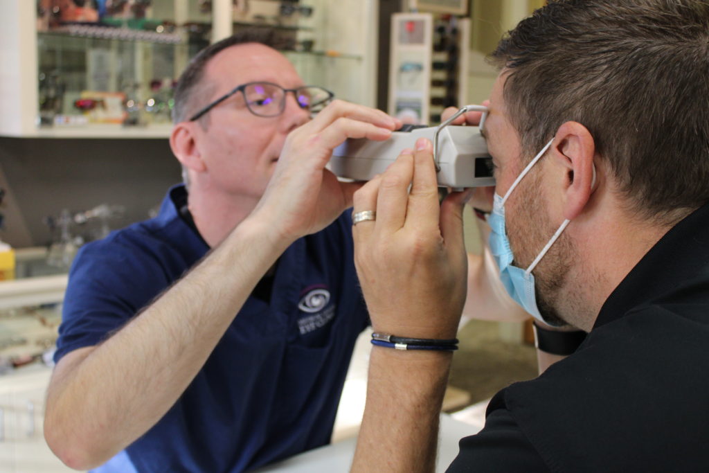
Each of our physicians has two assistants with them during the typical work day: a tech, and a writer, or scribe. The tech takes each patient to an exam room for their initial workup. The tech’s work includes reviewing a patient’s medical history, medications, and any complaints that the patient may have experienced. They also check the patient’s visual acuity and peripheral vision during each exam. Depending on the patient’s needs, the tech will also check their depth perception, color vision, or perform a dry eye test. This gets the patient ready for the doctor’s exam and helps the office flow efficiently.
The effects of dilation include trouble focusing up close and some light sensitivity. Once the pupils are fully dilated, the doctor uses the slit lamp microscope and different magnifying lenses to look at the eyes from front to back. During this examination is where the doctor would detect any conditions that may or may not need treatment. Some common ocular conditions are cataracts, diabetic retinopathy, macular degeneration, and glaucoma. Most of these diagnoses are initially made based on what the doctor sees during the exam. However, there are many tests we can perform to verify any findings that allow the doctor to further evaluate and manage each patient’s individual needs.
OCT – A SPECTRALIS® OCT examination provides information about the condition of the retinal layers. The examination can help identify early signs of disease in the retina and on the optic nerve head.
HVF/GVF – Humphrey Visual Field Analyzer 3/Goldmann Visual Field Analyzer – The Humphrey visual field test measures the entire area of peripheral vision that can be seen while the eye is focused on a central point. … The perception of these lights is charted and then compared to results of a healthy eye at the same age of the patient to determine if any damage has occurred
Fundus/Disc Photos -Nidek Non–Mydriatic Fundus Camera – This test is often done in addition to a routine eye exam to screen for eye diseases. Your eye doctor may also order it if you have a condition that affects your blood vessels, such as high blood pressure or diabetes. Ophthalmoscopy may also be called funduscopy or retinal examination.
Atlas 9000 Corneal Topography – Corneal topography is a computer assisted diagnostic tool that creates a three-dimensional map of the surface curvature of the cornea. … These details are used to diagnose, monitor, and treat various eye conditions. They are also used in fitting contact lenses and for planning surgery, including laser vision correction.
Biometry – Our Lenstar captures axial dimensions of all the human eye’s optical structures, from the cornea to the retina. This machine also measures corneal curvature, white-to-white, specific measurements for planning surgery, including laser vision correction, lens options and more.
OCT images are used to help diagnose diseases of the back of the eye: the retina, macula, and optic nerve. These images can show changes caused by macular degeneration, glaucoma, diabetic retinopathy, and many other conditions. The machine acts like a camera but can give us scans through the different layers of the retina to show things that may not be fully visible on an exam by the doctor. These images are typically taken once a year in patients already diagnosed with one of the above mentioned diseases.
Pachymetry – is a simple, painless test to measure the thickness of your cornea — the clear window at the front of the eye. A probe called a pachymeter is gently placed on the front of the eye (the cornea) to measure its thickness.
Sometimes the doctors will want to perform gonioscopy to clearly evaluate the angles at the front of the eye to determine if the drainage system is working correctly, which can also affect intraocular pressure. This test is performed by the doctor at the slit lamp microscope with a special mirrored lens that is held to the front of the eye. Gonioscopy is usually done once a year on patients with glaucoma, but can be done more frequently at the doctor’s discretion.
Dry Eye Testing: We start with a Tear Lab to determine the concentration of tears that your eyes produce. We also perform a Inflammadry test. Inflammadry is a rapid result eye test that detects inflammation due to high levels of MMP-9 in tears. We may also perform Fundus Retinal photos to record the condition of the interior surface of your eye. This allows us to document eye disorders and how they change inside your eye.
Cataracts
By the age of 80, more than half of Americans will have been diagnosed with cataracts or undergone surgery to have them removed. Cataract surgery is one of the most common surgeries in the United States. It is very simple and requires virtually no recovery for most patients. During the surgery, the doctor will remove the natural lens inside the eye that is affected by the cataract, and replace it with a man-made lens. We accept assignment insurance coverage.
If a patient is interested in scheduling cataract surgery, they schedule an appointment with our surgery coordinator. The patient will be scheduled for a pre-op appointment to obtain measurements and go over instructions for their surgery. These measurements include ultrasound or manual keratometry, corneal topography, and A-Scan. These measurements together help the doctor make accurate decisions regarding the lens he will use to replace the natural lens in the eye affected by the cataract.
An ophthalmologist will also do various testing to screen for glaucoma, which is a disease that usually involves increased intraocular pressure (pressure from the fluid in the eye), and can cause damage to the optic nerve and vision loss. Eye pressure is measured with a tonometer, a probe-like device that measures the eye. In patients with glaucoma, it also helps to obtain visual field measurements, OCT images, pachymetry, and gonioscopy.
With all the testing we have available, your ophthalmologists in Salem can catch most conditions early and treat accordingly. The tests are all easy on the patient and usually very quick. Due to the many conditions that can affect your eyes and vision in various ways, it is very important to have an ophthalmologist that can help make sure your eyes are healthy now and into the future.